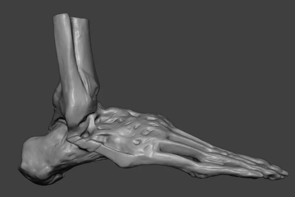
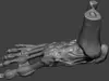
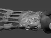
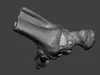
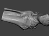
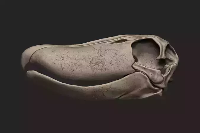
A high-fidelity 3D anatomical model of the human foot, presented in a premium écorché style. This model offers a comprehensive view of the complex biomechanical architecture of the lower limb's distal extremity.
Key Anatomical Features:
- Osteology: Accurate representation of the Calcaneus (heel), Talus, Tarsals, Metatarsals, and Phalanges.
- Myology & Syndesmology: Detailed visualization of the extensor and flexor tendons, dorsal interosseous muscles, and the complex ligamentous structures forming the foot arches.
- Plantar Fascia: Exceptional rendering of the plantar aponeurosis on the sole.
- Cross-Section: The proximal end shows a clean anatomical cut of the distal Tibia and Fibula.
- Digital Quality: Processed from a high-resolution 3D scan with professional retopology (clean quad-dominant mesh).
Perfect for orthopedic visualization, physical therapy education, podiatry study, and high-end digital character reference.