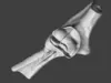
Human Elbow Joint: Bones and Ligaments (Syndesmology Model)
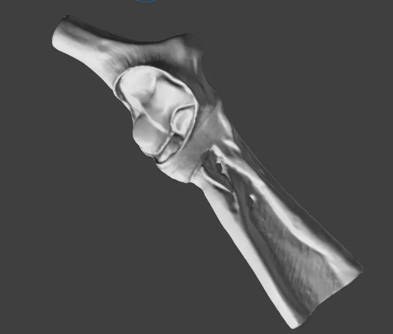
A high-fidelity 3D anatomical model of the human elbow joint. This model offers a detailed look at the complex interaction between the distal humerus, proximal ulna, and radius, along with the essential ligamentous apparatus.
Key Anatomical Features:
- Bony Landmarks: Clearly defined trochlea and capitulum of the humerus, the olecranon process of the ulna, and the radial head.
- Ligaments: Visualization of the collateral ligaments (radial and ulnar) and the annular ligament, which are critical for joint stability.
- Articular Surfaces: Smooth, professionally processed surfaces that demonstrate how the hinge and pivot components of the elbow function together.
Generated from a high-resolution 3D scan and professionally retopologized for clean quad-dominant topology. Perfect for orthopedic studies, physical therapy education, and high-end CGI.