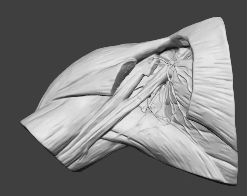
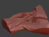
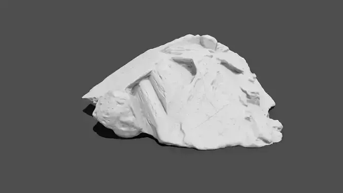
A high-fidelity, medical-grade 3D model of the human axillary region (axilla), meticulously reconstructed from a high-resolution 3D scan of an actual anatomical specimen. This écorché model provides an exceptional topographic view of the neurovascular bundle and the surrounding musculature.This asset is designed for professionals requiring superior anatomical accuracy for medical visualization, surgical planning, or high-end character technical directing (TD).
Key Anatomical FeaturesBrachial Plexus (Plexus Brachialis): Highly detailed rendering of the nerve cords and terminal branches, including the Median, Ulnar, Radial, and Musculocutaneous nerves.Vascular System: Accurately placed Axillary Artery and Axillary Vein.Musculature: Features the Pectoralis Major and Minor (sectioned to reveal depth), Coracobrachialis, and the short head of the Biceps Brachii.Topography: Clear visualization of the axillary walls and the pathway of nerves passing from the neck/chest to the upper limb.
Technical SpecificationsGeometry: Clean, quad-dominant topology (optimized via professional retopology).Mesh: Manifold, watertight, and optimized for high-end rendering and medical CGI.Detailing: High-poly surface preserves organic muscle fiber directions and fascia textures.Scale: 1:1 Human Life-Size (Metric units).Formats Included: .STL (Print-ready), .OBJ, .FBX (Digital use).
Use CasesMedical Education: Advanced study of regional anesthesia and surgical approaches to the axilla.CGI & VFX: High-quality source for baking maps onto character models or for anatomical reference.3D Printing: Ideal for medical museums or classroom aids (Resin/SLA printing recommended for nerve detail).