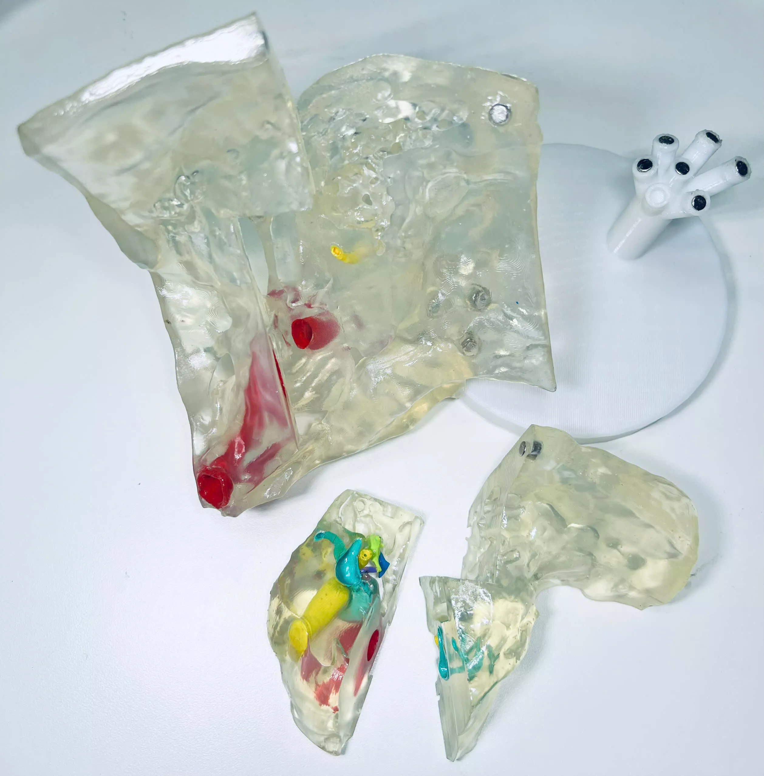
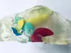
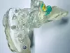
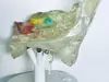
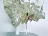
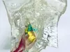
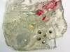
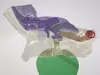
The development process of this model was published in MedPharmRes journal. The corresponding paper is included in the ZIP file.Explore highly detailed temporal bone anatomy with this 3D printable model, reconstructed from a real patient CT scan. Designed for mastoidectomy drilling practice, surgical simulation, and medical education, this model provides exceptional anatomical accuracy of the middle and inner ear.Key Features
- Two Versions Included: 3-Part Model (Bone 1, Bone 2, Bone 3) – optimized for easy 3D printing
- Intact Temporal Bone (1-piece) – ideal for mastoidectomy drilling practiceHighly Detailed Anatomy with STL file for each structure:
- Ossicles: malleus, incus, stapes
- Cochlea & vestibule
- Semicircular canals (labyrinth)
- Facial nerve (CN VII – segments 1, 2, 3)
- Vestibulocochlear nerve (CN VIII)
- Internal carotid artery
- Mastoid air cellsReal Patient CT-Based Reconstruction. Ensures true anatomical accuracy for clinical learningMulti-Part & Multi-Material Ready: 8 separate STL components for detailed color printingCompatible with multi-color / resin / FDM printingFormats included: STL, OBJ, MTL, GLB, FBXOrganized in a ZIP folderPre-merged parts (Bone 1–3) for easy printingSeparate structures for advanced customization3D Printing Recommendations
- Transparent resin model: print at 200% scale for visualization
- Drilling model (1-piece): print at 100% scale
- Recommended magnets: 5×3 mm or 6×4 mm neodymium magnets (for assembly versions)Ideal For:
- ENT surgeons & residents (mastoidectomy training)
- Radiologists & anatomy educators
- Medical students learning temporal bone anatomy
- Simulation labs & teaching hospitals