DESCRIPTION

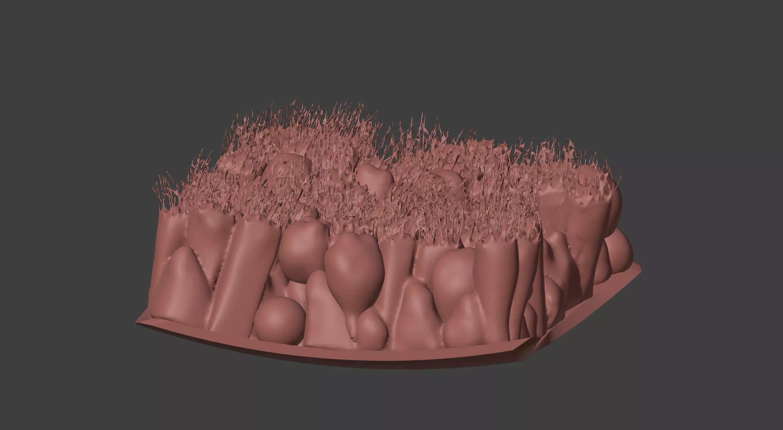
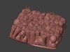
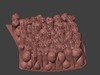
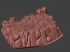
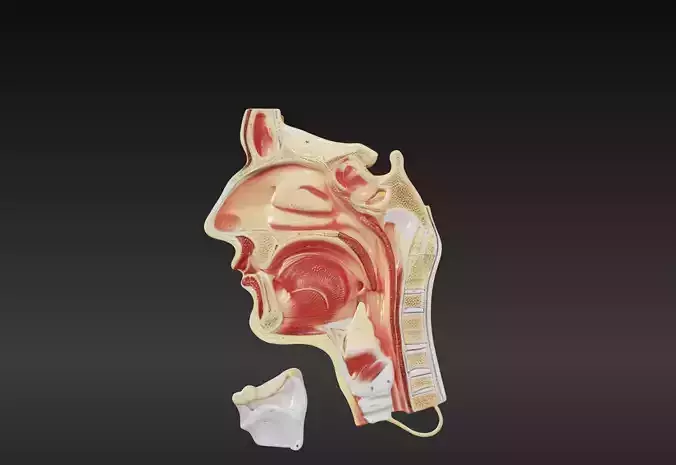
This 3D model of the epithelium of the trachea provides a highly detailed and anatomically accurate representation of the tracheal wall and its cellular structure. It highlights the pseudostratified ciliated columnar epithelium, goblet cells, and basal cells, showcasing their roles in mucus production and debris removal. The model also illustrates the submucosa, hyaline cartilage rings, and smooth muscle layers, offering insights into the structural support and flexibility of the trachea.

Designed for educational and clinical purposes, this model is an invaluable tool for studying respiratory anatomy, understanding airway function, and visualizing conditions such as tracheitis, inflammation, and mucus buildup. Ideal for pulmonologists, educators, and students, it supports teaching, training, and patient education by providing a hands-on approach to exploring the structure and function of the tracheal epithelium and respiratory system.

REVIEWS & COMMENTS

See what other buyers think about this model - real feedback on quality,
accuracy, and usability.
There are no reviews or comments yet. Please be the first one to write it.

3D Model of the Epithelium of the Trachea 3D print model

Royalty Free License
Hire
Like this model to show appreciation to the designer.
See how many times this model was viewed.
Share this model to support the designer and boost their visibility.
File formats
STL
Stereolithography<br />File Size: 39.2 MB
OBJ
OBJ | 2 files<br />File Size: 64.5 MB
FBX
Autodesk FBX<br />File Size: 28 MB
DAE
Collada<br />File Size: 189 MB
BLEND
Blender<br />File Size: 85.1 MB
GLTF
glTF<br />File Size: 36.5 MB
Provided by designer
Information and details shared directly by the model's designer.
3D printing
Indicates whether the designer marked this model as suitable for 3D printing.
Model is prepared for 3D printing
The designer indicates this model is prepared for physical 3D printing and includes printable file formats.
Split into parts
Units
Publish date
Model ID
Chat