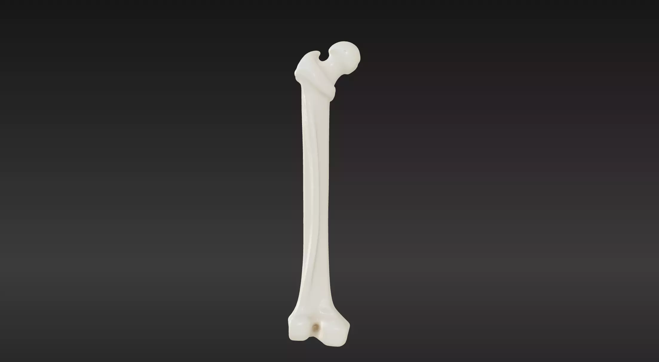
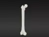
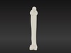
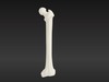
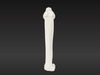
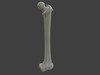
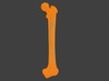
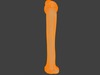
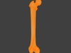
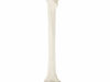
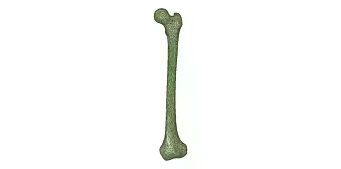
This 3D model of the femur bone is a highly detailed and anatomically accurate representation of the human thigh bone. Designed for educational, medical, and professional purposes, it showcases the femur's distinct features, including:
- Proximal End: Detailed structures of the femoral head, neck, and greater and lesser trochanters.
- Shaft: Accurately shaped diaphysis with realistic curvature and surface textures.
- Distal End: Clearly defined medial and lateral condyles, intercondylar notch, and epicondyles.
The model is optimized for 3D printing and is ideal for use in:
- Medical Education: Demonstrate bone anatomy, structure, and landmarks.
- Orthopedic Studies: Analyze and simulate fractures, prosthetics, and surgical planning.
- Art and Design: Use as a reference for artistic projects requiring realistic anatomy.
The file is available in multiple formats (e.g., STL, OBJ) for compatibility with various 3D printers and software. Whether you're a medical professional, educator, or 3D enthusiast, this femur bone model offers unmatched precision and usability.