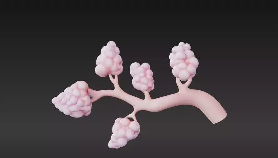
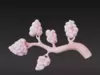
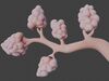
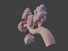
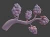
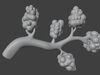
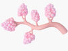
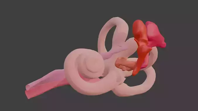
This 3D model of lactiferous duct anatomy is an accurate and detailed representation of the intricate ductal system within the . Designed for educational, medical, and research applications, it provides a clear visualization of the structures responsible for milk transport during lactation.
Key features:
- Detailed depiction of the lactiferous ducts, including their branching structure leading from the lobules to the .
- Highlights the lobules (milk-producing glands) and their connection to the lactiferous ducts.
- Clear visualization of the lactiferous sinuses near the , where milk is stored before secretion.
- Demonstrates the relationship between the ducts, surrounding tissue, and key anatomical landmarks such as the and areola.
- Realistic proportions and textures to replicate the internal anatomy of the .
Applications:
- Medical education to teach the anatomy and function of the lactiferous ducts and their role in breastfeeding.
- Research in health, including studies of conditions like mastitis, ductal carcinoma, and other duct-related pathologies.
- Artistic and scientific reference for creating accurate anatomical illustrations or 3D visualizations.
The model is optimized for 3D printing and digital use, provided in commonly used formats such as STL and OBJ, ensuring compatibility with various software and 3D printers. This lactiferous duct anatomy model is an invaluable resource for educators, researchers, and designers seeking a precise representation of this critical structure.