1/7
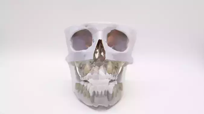
3D Printed Palatal Expansion Model from Patient DICOM Data
This model was created directly from segmented DICOM imaging of a young patient requiring palatal expansion. By transforming radiographic data into a physical 3D print, we’re able to precisely replicate the patient’s internal anatomy, not just the external form.
Unlike traditional dental typodonts, this model demonstrates the true bone structure, midpalatal suture, and surrounding anatomy as captured in the original scan. The level of internal detail provides clinicians and students with a more accurate understanding of how palatal expansion impacts skeletal and dental structures in growing patients.
Because the model is derived from real patient data, it highlights variations in anatomy that are critical for diagnosis, treatment planning, and training. This allows orthodontic teams to visualize and practice procedures on a physical replica before applying them clinically, enhancing both safety and educational value.
REVIEWS & COMMENTS
accuracy, and usability.


