DESCRIPTION

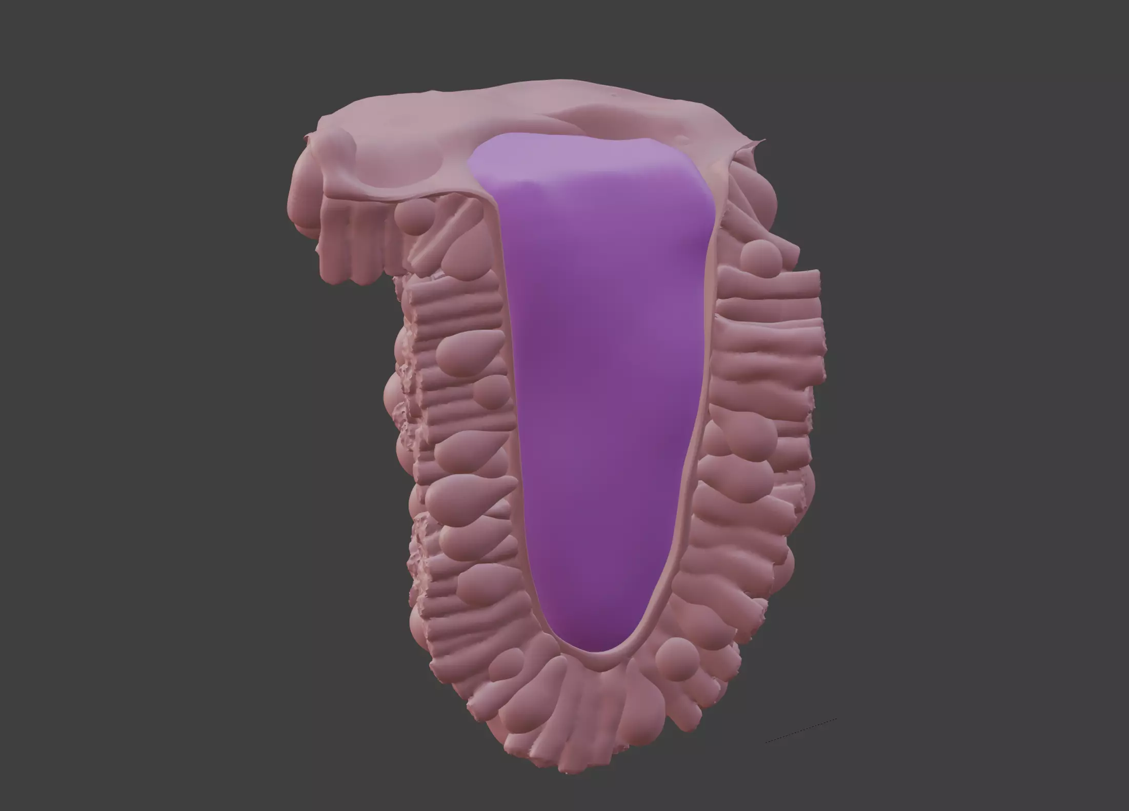
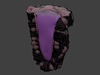
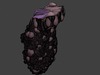
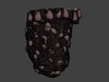
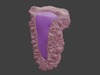
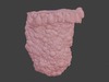
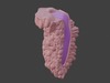
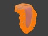
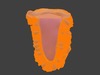
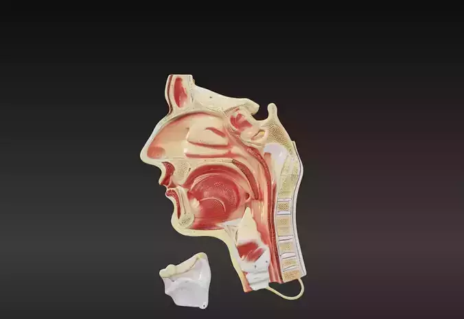
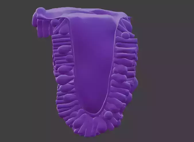
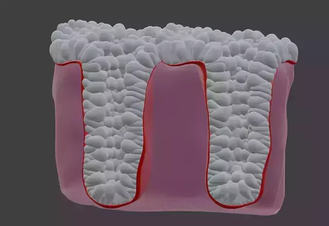
This 3D model of the epithelium of the duodenum provides a highly detailed and anatomically accurate representation of the structural layers of the duodenal wall. It highlights the simple columnar epithelium with microvilli, goblet cells, and intestinal villi, which increase the surface area for nutrient absorption.

The model also illustrates key features such as crypts of Lieberkühn, Brunner’s glands in the submucosa, and smooth muscle layers, demonstrating the duodenum’s role in digestion and absorption.

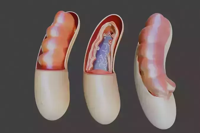
Designed for educational and clinical purposes, this model is an invaluable tool for studying gastrointestinal anatomy, understanding digestive processes, and visualizing conditions such as malabsorption syndromes, ulcers, and inflammation.

Ideal for gastroenterologists, educators, and students, it supports teaching, training, and patient education by providing a hands-on approach to exploring the structure and function of the duodenal epithelium.

REVIEWS & COMMENTS

See what other buyers think about this model - real feedback on quality,
accuracy, and usability.
There are no reviews or comments yet. Please be the first one to write it.

3D Model of the Epithelium of the Duodenum 3D print model

Royalty Free License
Hire
Like this model to show appreciation to the designer.
See how many times this model was viewed.
Share this model to support the designer and boost their visibility.
File formats
STL
Stereolithography<br />File Size: 34.5 MB
OBJ
OBJ | 2 files<br />File Size: 52.6 MB
BLEND
Blender<br />File Size: 54.1 MB
FBX
Autodesk FBX<br />File Size: 20.8 MB
DAE
Collada<br />File Size: 135 MB
GLTF
glTF<br />File Size: 24.8 MB
Provided by designer
Information and details shared directly by the model's designer.
3D printing
Indicates whether the designer marked this model as suitable for 3D printing.
Model is prepared for 3D printing
The designer indicates this model is prepared for physical 3D printing and includes printable file formats.
Split into parts
Units
Publish date
Model ID
Chat