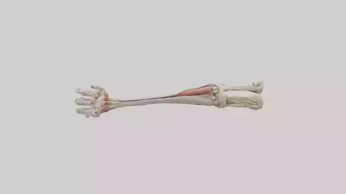
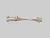
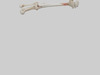
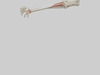
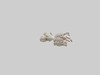
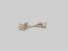
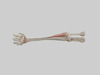
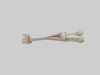
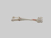
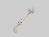
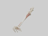
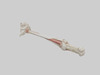
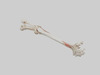
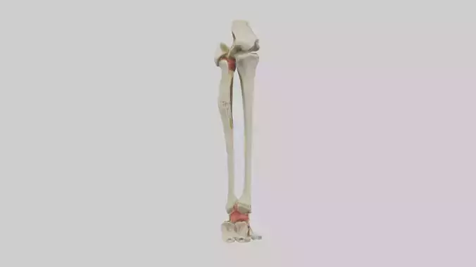
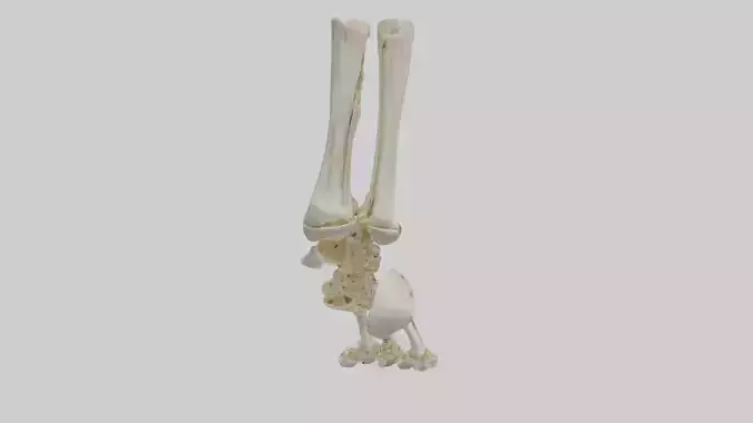
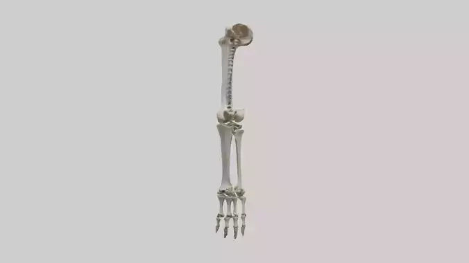
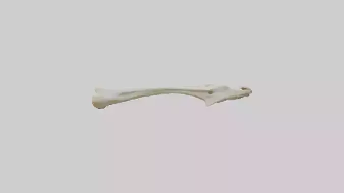
İnsan Radius ve Ulna Modeli'nin kısa 3D modeli. İnsan iskeletinin ön kolunda bulunan iki uzun kemik olan radius ve ulna'nın 3D temsilini tasvir eder. Radius tipik olarak ikisinin daha kısasıdır ve ön kolun başparmak tarafında bulunur. Ulna daha uzundur ve serçe parmağı tarafında bulunur. Model, radyal baş, radyal tüberkül, ulnar baş, olekranon çıkıntısı ve koranoid çıkıntı gibi spesifik kemiksi yapıları içeren her bir kemiğin anatomik özelliklerini doğru bir şekilde göstermelidir. Model, kemikleri birbirlerine göre doğal pozisyonlarında eklemlenmiş olarak, proksimal ve distal radioulnar eklemleri ve aralarındaki interosseöz membran boşluğunu göstererek sunulabilir. Ayrıntı seviyesi, anatomi ve biyoloji alanlarında eğitim amaçları için yeterli olmalıdır. Stil muhtemelen gerçekçi ve anatomik olarak doğru olacaktır. Bu model, tıp öğrencileri, eğitimciler, sağlık profesyonelleri ve insan anatomisi üzerine çalışan herkes için uygundur. FBX, OBJ, STL, GLB, USDZ ve BLEND formatlarında mevcuttur.
Türkçe Anahtar Kelimeler:
İnsan Radius ve Ulna Modeli, 3D model, radius, ulna, ön kol, kemik, iskelet, anatomi, biyoloji, tıp, eğitim, FBX, OBJ, STL, GLB
İngilizce Açıklama:
Concise 3D model of the Human Radius and Ulna. Depicts a 3D representation of the two long bones in the forearm of the human skeleton: the radius and the ulna. The radius is typically the shorter of the two and is located on the thumb side of the forearm. The ulna is longer and located on the pinky side. The model should accurately show the anatomical features of each bone, including the diaphysis (shaft), epiphyses (ends), and specific bony landmarks like the radial head, radial tuberosity, ulnar head, olecranon process, and coronoid process. The model might show the bones articulated in their natural position relative to each other, demonstrating the proximal and distal radioulnar joints and the interosseous membrane space between them. The level of detail should be sufficient for educational purposes in anatomy and biology. The style would likely be realistic and anatomically accurate. Suitable for medical students, educators, healthcare professionals, and anyone studying human anatomy. Available in FBX, OBJ, STL, GLB, USDZ, and BLEND formats.
İngilizce Anahtar Kelimeler:
Human Radius and Ulna Model, 3d model, radius, ulna, forearm, bone, skeleton, anatomy, biology, medical, education, FBX, OBJ, STL, GLB
Almanca Beschreibung:
Kurzes 3D-Modell der menschlichen Speiche und Elle. Zeigt eine 3D-Darstellung der beiden langen Knochen im Unterarm des menschlichen Skeletts: Radius (Speiche) und Ulna (Elle). Der Radius ist typischerweise der kürzere der beiden und befindet sich auf der Daumenseite des Unterarms. Die Ulna ist länger und befindet sich auf der Kleinfingerseite. Das Modell sollte die anatomischen Merkmale jedes Knochens genau darstellen, einschließlich der Diaphyse (Schaft), der Epiphysen (Enden) und spezifischer knöcherner Landmarken wie des Radiusköpfchens, der Tuberositas radii, des Ulnaköpfchens, des Olecranon und des Processus coronoideus. Das Modell könnte die Knochen in ihrer natürlichen Position zueinander artikuliert zeigen und die proximalen und distalen Radioulnargelenke sowie den Raum der Membrana interossea zwischen ihnen darstellen. Der Detaillierungsgrad sollte für Bildungszwecke in Anatomie und Biologie ausreichend sein. Der Stil wäre wahrscheinlich realistisch und anatomisch korrekt. Geeignet für Medizinstudenten, Pädagogen, Angehörige der Gesundheitsberufe und alle, die menschliche Anatomie studieren. Verfügbar in den Formaten FBX, OBJ, STL, GLB, USDZ und BLEND.
Almanca Schlüsselwörter:
Menschliches Radius und Ulna Modell, 3D Modell, Radius, Ulna, Unterarm, Knochen, Skelett, Anatomie, Biologie, Medizin, Bildung, FBX, OBJ, STL, GLB
Lehcze Opis:
Krótki model 3D Kości Promieniowej i Łokciowej Człowieka. Przedstawia trójwymiarową reprezentację dwóch długich kości przedramienia szkieletu człowieka: kości promieniowej (radius) i kości łokciowej (ulna). Kość promieniowa jest zazwyczaj krótsza i znajduje się po stronie kciuka przedramienia. Kość łokciowa jest dłuższa i znajduje się po stronie małego palca. Model powinien dokładnie pokazywać cechy anatomiczne każdej kości, w tym trzon (diaphysis), nasady (epiphyses) i specyficzne punkty kostne, takie jak głowa kości promieniowej, guzowatość kości promieniowej, głowa kości łokciowej, wyrostek łokciowy i wyrostek dziobiasty. Model może przedstawiać kości połączone w ich naturalnym położeniu względem siebie, ukazując stawy promieniowo-łokciowe bliższy i dalszy oraz przestrzeń błony międzykostnej między nimi. Poziom szczegółowości powinien być wystarczający do celów edukacyjnych w anatomii i biologii. Styl prawdopodobnie będzie realistyczny i anatomicznie dokładny. Nadaje się dla studentów medycyny, pedagogów, pracowników służby zdrowia i wszystkich studiujących anatomię człowieka. Dostępny w formatach FBX, OBJ, STL, GLB, USDZ i BLEND.
Lehcze Słowa kluczowe:
model Kości Promieniowej i Łokciowej Człowieka, model 3D, kość promieniowa, kość łokciowa, przedramię, kość, szkielet, anatomia, biologia, medycyna, edukacja, FBX, OBJ, STL, GLB
Arapça وصف:
نموذج ثلاثي الأبعاد موجز لعظم الكعبرة والزند البشري. يصور نموذجًا ثلاثي الأبعاد للعظمين الطويلين في الساعد من الهيكل العظمي البشري: الكعبرة والزند. الكعبرة هي عادةً الأقصر بينهما وتقع على جانب الإبهام من الساعد. الزند أطول ويقع على جانب الخنصر. يجب أن يُظهر النموذج بدقة الميزات التشريحية لكل عظم، بما في ذلك الديابيز (الجسم) والنهايات (الأطراف) والمعالم العظمية المحددة مثل رأس الكعبرة وحدبة الكعبرة ورأس الزند والناتئ المرفقي والناتئ الإكليلي. قد يُظهر النموذج العظام متمفصلة في وضعها الطبيعي بالنسبة لبعضها البعض، مما يدل على المفصلين الكعبري الزندي القريب والبعيد والفضاء الغشائي بينهما. يجب أن يكون مستوى التفاصيل كافيًا للأغراض التعليمية في علم التشريح وعلم الأحياء. من المحتمل أن يكون الأسلوب واقعيًا ودقيقًا من الناحية التشريحية. مناسب لطلاب الطب والمعلمين والمتخصصين في الرعاية الصحية وأي شخص يدرس علم التشريح البشري. متوفر بتنسيقات FBX و OBJ و STL و GLB و USDZ و BLEND.
Arapça كلمات مفتاحية:
نموذج عظم الكعبرة والزند البشري, نموذج ثلاثي الأبعاد, كعبرة, زند, ساعد, عظم, هيكل عظمي, تشريح, علم الأحياء, طبي, تعليم, FBX, OBJ, STL, GLB
Çincese 描述:
人类桡骨和尺骨的简洁 3D 模型。描绘了人类骨骼前臂中的两根长骨:桡骨和尺骨的 3D 模型。桡骨通常较短,位于前臂的拇指侧。尺骨较长,位于小指侧。该模型应准确显示每根骨骼的解剖特征,包括骨干、骨骺以及特定的骨性标志,如桡骨头、桡骨粗隆、尺骨头、鹰嘴突和冠状突。该模型可以显示骨骼以其相对于彼此的自然位置进行关节连接,展示近端和远端桡尺关节以及它们之间的骨间膜间隙。细节程度应足以满足解剖学和生物学的教育目的。风格很可能是写实的并且在解剖学上是准确的。适用于医学生、教育工作者、医疗保健专业人员以及任何学习人体解剖学的人。提供 FBX、OBJ、STL、GLB、USDZ 和 BLEND 格式。
Çincese 关键词:
人类桡骨和尺骨模型, 3D 模型, 桡骨, 尺骨, 前臂, 骨骼, 骨骼, 解剖学, 生物学, 医学, 教育, FBX, OBJ, STL, GLB
Korece 설명:
인간 요골 및 척골 모델의 간결한 3D 모델입니다. 인간 골격의 팔뚝에 있는 두 개의 긴 뼈인 요골과 척골의 3D 표현을 묘사합니다. 요골은 일반적으로 두 개 중 더 짧고 팔뚝의 엄지손가락 쪽에 위치합니다. 척골은 더 길고 새끼손가락 쪽에 위치합니다. 모델은 요골두, 요골 거친면, 척골두, 팔꿈치머리돌기 및 갈고리돌기와 같은 특정 뼈 구조를 포함하여 각 뼈의 해부학적 특징을 정확하게 보여주어야 합니다. 모델은 뼈가 서로에 대한 자연스러운 위치로 연결되어 근위 및 원위 요척관절과 그 사이의 뼈사이막 공간을 보여줄 수 있습니다. 세부 수준은 해부학 및 생물학 교육 목적에 충분해야 합니다. 스타일은 사실적이고 해부학적으로 정확할 가능성이 높습니다. 의대생, 교육자, 의료 전문가 및 인체 해부학을 공부하는 모든 사람에게 적합합니다. FBX, OBJ, STL, GLB, USDZ 및 BLEND 형식으로 제공됩니다.
Korece 키워드:
인간 요골 및 척골 모델, 3D 모델, 요골, 척골, 팔뚝, 뼈, 골격, 해부학, 생물학, 의학, 교육, FBX, OBJ,